
Dale Dubin’s Rapid Interpretation of EKGs is a renowned guide for mastering electrocardiogram analysis․ It offers a comprehensive, interactive approach to understanding EKG principles, recording techniques, and clinical applications, making it an invaluable resource for both medical students and professionals․
1․1 Overview of Dale Dubin’s Work
Dale Dubin, a prominent figure in medical education, authored the acclaimed Rapid Interpretation of EKGs, a comprehensive guide for understanding electrocardiography․ His work emphasizes a structured, interactive approach, making complex concepts accessible․ The book covers essential topics like basic EKG principles, heart rate, rhythm, blocks, axis, hypertrophy, and infarction․ Dubin’s methodical teaching style has made his resources indispensable for medical professionals and students worldwide, ensuring accurate and efficient EKG interpretation skills․
1․2 Importance of EKG Interpretation in Medical Practice
EKG interpretation is a cornerstone of medical practice, enabling early detection of cardiac abnormalities․ It guides diagnosis, treatment, and monitoring of heart conditions․ Accurate EKG reading helps identify arrhythmias, hypertrophy, and infarction, crucial for timely interventions․ Dubin’s work emphasizes its role in improving patient outcomes, making it a fundamental skill for healthcare professionals․ Its non-invasive nature provides immediate insights, essential in emergency settings, ensuring precise and life-saving decisions․
Key Features of “Rapid Interpretation of EKGs”
Rapid Interpretation of EKGs offers an interactive, structured approach to learning EKG analysis․ It includes comprehensive coverage of basic principles, recording techniques, and clinical applications, making it a valuable resource for medical professionals;
2․1 Structure and Content of the Book
Rapid Interpretation of EKGs is structured to provide a logical progression from basic principles to advanced concepts․ The book is divided into sections covering EKG recording, the autonomic nervous system, heart rate, rhythm, arrhythmias, blocks, electrical axis, hypertrophy, myocardial infarction, and miscellaneous effects․ It includes detailed chapters on rate and rhythm analysis, axis determination, and the impact of pathological conditions․ The content is supported by case studies, quick reference sheets, and practical tips, making it a comprehensive guide for both learners and practitioners․
2․2 Unique Approach to EKG Learning
Dale Dubin’s Rapid Interpretation of EKGs employs an interactive, programmed learning style that engages readers actively․ The book uses a step-by-step approach, breaking down complex concepts into manageable parts․ It incorporates practical exercises, case studies, and tracing examples to reinforce learning․ Dubin’s method emphasizes pattern recognition and correlation with clinical scenarios, making it easier to apply knowledge in real-world situations․ This unique blend of theory and practice sets the book apart as an effective tool for mastering EKG interpretation․

Basic Principles of EKG
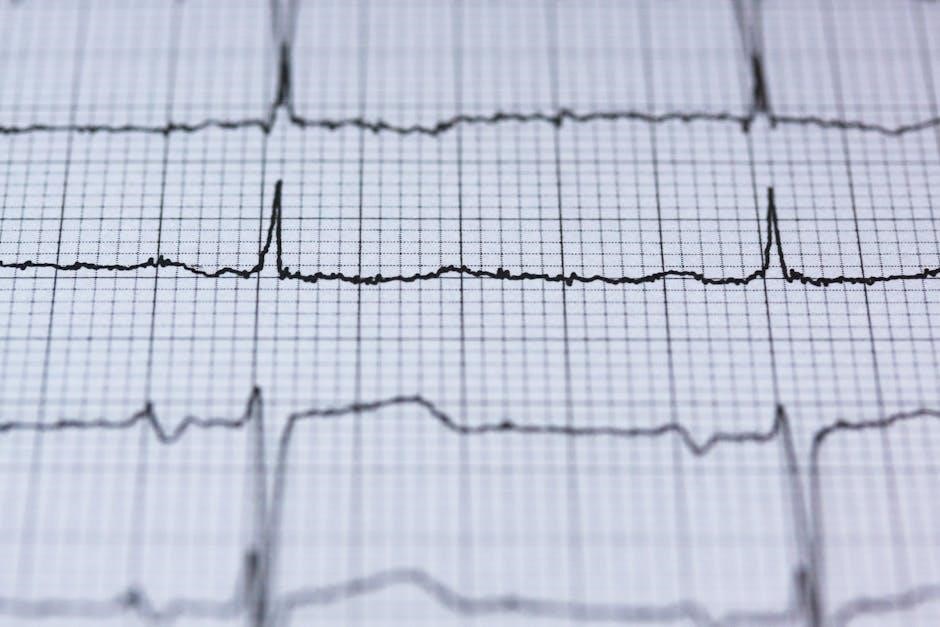
Dale Dubin’s guide explains the fundamental science behind EKG recordings, focusing on electrical heart activity and its graphical representation․ It covers essential components like P, QRS, and T waves, ensuring a solid foundation for interpretation․

3․1 The Science Behind EKG Recordings
EKG recordings capture the electrical activity of the heart, translating muscle depolarization into measurable waveforms․ The P wave represents atrial depolarization, while the QRS complex signifies ventricular depolarization․ The T wave reflects ventricular repolarization․ These components provide insights into heart function, rhythm, and potential abnormalities․ Dubin’s guide emphasizes understanding the physiological basis of these recordings, enabling accurate interpretation of heart health and dysfunction․
3․2 Components of an EKG Tracing
An EKG tracing consists of distinct components: the P wave, QRS complex, and T wave․ The P wave represents atrial depolarization, while the QRS complex signifies ventricular depolarization․ The T wave reflects ventricular repolarization․ Additional elements include the PR interval (atrioventricular node delay) and QT interval (ventricular depolarization and repolarization)․ These components provide critical data for assessing heart rhythm, conduction, and potential abnormalities, as detailed in Dubin’s guide for precise EKG interpretation․

Recording the EKG
Recording an EKG involves placing electrodes on the skin to capture heart activity, using standard limb and chest leads․ Proper skin preparation and a calm environment ensure clear tracings․
4․1 Standard Limb and Chest Leads
Standard limb leads (I, II, III) and chest leads (V1-V6) are essential for capturing a comprehensive EKG․ Limb leads provide a view of the heart from different angles, while chest leads offer a direct view of the ventricles․ Together, they help identify electrical activity, axis deviation, and potential abnormalities․ Proper placement of these electrodes ensures accurate recordings, as emphasized in Dubin’s guide, which also covers troubleshooting common issues that may arise during the recording process to ensure clear and interpretable tracings․
4․2 Proper Electrode Placement
Proper electrode placement is critical for accurate EKG recordings․ Chest electrodes (V1-V6) are placed across the sternum and ribcage, while limb electrodes are positioned on the arms and legs․ Correct placement ensures clear signals and minimizes interference․ Dubin’s guide emphasizes avoiding movement and ensuring good skin contact․ Improper placement can lead to artifacts or poor trace quality, making interpretation challenging․ Adherence to standardized positions is vital for reliable and consistent EKG results, as outlined in the guide to help users achieve optimal recordings every time․

The Autonomic Nervous System and EKG
The autonomic nervous system influences heart rate and rhythm, with sympathetic activity increasing heart rate and parasympathetic activity promoting relaxation, both visible on EKG tracings․
5․1 Sympathetic and Parasympathetic Effects
The sympathetic nervous system increases heart rate and contractility, while the parasympathetic system promotes relaxation and reduces heart rate․ These effects are visible on EKG tracings, with sympathetic activity often causing an increased heart rate and shortened PR interval, and parasympathetic activity leading to a decreased heart rate and prolonged PR interval․ Understanding these interactions is crucial for interpreting EKG changes in various clinical conditions, as described in Dubin’s guide․
5․2 Catecholamines and Heart Rate
Catecholamines, such as adrenaline and noradrenaline, significantly influence heart rate by stimulating beta-adrenergic receptors․ This increases sinoatrial node activity, leading to a faster heart rate and shortened PR interval on EKG․ Dubin’s guide explains how these physiological responses are reflected in EKG tracings, aiding in the identification of conditions like hyperadrenergic states or stress-induced tachycardia․ Understanding catecholamine effects is essential for accurate EKG interpretation in clinical settings․
Heart Rate and Rhythm
Dubin’s guide explains heart rate calculation and rhythm analysis, distinguishing normal sinus rhythms from abnormal patterns like tachycardia or bradycardia, essential for accurate EKG interpretation․
6․1 Calculating Heart Rate from EKG
Dubin’s guide provides clear methods for calculating heart rate from EKG tracings, such as the 1500 method and measuring RR intervals․ Practical examples and step-by-step instructions help learners master accurate heart rate determination, essential for identifying normal sinus rhythms and detecting conditions like tachycardia or bradycardia; This section emphasizes the importance of precise calculations in clinical decision-making, ensuring reliable EKG interpretation skills․
6․2 Normal and Abnormal Rhythms
Dubin’s guide thoroughly explains normal sinus rhythms and their variations, such as sinus arrhythmia․ It also details abnormal rhythms, including atrial fibrillation, flutter, and ventricular ectopics․ Clear examples and diagrams help differentiate between benign and pathological conditions․ The section emphasizes recognizing P-wave patterns, PR intervals, and QRS complexes to accurately identify rhythm disturbances․ This knowledge is crucial for diagnosing arrhythmias and understanding their clinical implications, making it an essential skill for healthcare professionals and students alike․
Arrhythmias of Focal Origin
Arrhythmias of focal origin arise from specific heart regions, such as atrial or ventricular ectopics, and are crucial for accurate EKG interpretation and diagnosis․
7․1 Atrial and Junctional Arrhythmias
Atrial and junctional arrhythmias originate from specific regions in the heart․ Atrial arrhythmias, such as premature atrial contractions and atrial fibrillation, are common and often benign but can indicate underlying conditions․ Junctional arrhythmias, like junctional tachycardia, arise from the AV junction․ Dubin’s guide provides clear criteria for identifying these rhythms on EKG, emphasizing P-wave morphology and PR interval analysis․ His systematic approach helps differentiate between benign and pathological arrhythmias, ensuring accurate diagnosis and appropriate management․
7․2 Ventricular Ectopics and Tachycardia
Ventricular ectopics and tachycardia are arrhythmias originating in the ventricles․ Dubin’s guide details how to identify these on EKG, focusing on wide QRS complexes and T-wave discordance․ Ventricular ectopic beats are often benign but can signal underlying heart disease․ Ventricular tachycardia, characterized by three or more consecutive ectopics, may indicate serious conditions like ischemia or cardiomyopathy․ Dubin’s systematic approach helps differentiate these rhythms, emphasizing clinical correlation to determine their significance and appropriate management․

Blocks in EKG Interpretation
Dubin’s guide explains various EKG blocks, such as AV and Bundle Branch Blocks, and their identification through P-QRS-T interval analysis, aiding in accurate diagnosis and treatment plans․
8․1 AV and Bundle Branch Blocks
Dale Dubin’s guide thoroughly explains AV and Bundle Branch Blocks, focusing on their identification and implications․ AV blocks disrupt electrical signals between the atria and ventricles, while Bundle Branch Blocks affect ventricular conduction․ The EKG reveals characteristic patterns, such as prolonged PR intervals in AV blocks and widened QRS complexes in Bundle Branch Blocks․ Understanding these abnormalities is crucial for accurate diagnosis and treatment, as they often indicate underlying cardiac conditions requiring prompt medical attention․
8․2 Fascicular and Bifascicular Blocks
Fascicular Blocks involve the left anterior or posterior fascicle, altering EKG patterns with specific notching or axis deviations․ Bifascicular Blocks affect two fascicles, often presenting as left bundle branch block with left anterior fascicular block․ Dubin’s guide emphasizes identifying these patterns, as they may indicate progressive conduction disease․ Clinical correlation is crucial, as Bifascicular Blocks can precede complete heart block, necessitating prompt medical evaluation and potential pacing․ These insights are vital for accurate EKG interpretation and patient management․

Electrical Axis and Its Significance
The electrical axis in EKGs indicates the heart’s electrical impulse direction, aiding in diagnosing conditions like left or right axis deviation, crucial for accurate patient assessment․
9․1 Determining the Electrical Axis
Determining the electrical axis involves analyzing the limb leads to identify the direction of the heart’s electrical impulse․ Dubin’s method emphasizes calculating the net deflection in leads I, II, and III․ A normal axis ranges from -30° to +100°․ Deviations, such as left or right axis deviation, indicate potential cardiac conditions․ The P-wave axis is also assessed to confirm atrial depolarization patterns․ Accurate axis determination is crucial for diagnosing conditions like bundle branch blocks or ventricular hypertrophy, guiding further clinical evaluation and management․
9․2 Axis Deviation in Pathological Conditions
Axis deviation on an EKG can indicate underlying cardiac pathology․ Left axis deviation is often associated with left ventricular hypertrophy or bundle branch blocks, while right axis deviation may suggest right ventricular hypertrophy or pulmonary disease․ Dubin’s guide highlights how axis deviation aids in diagnosing conditions like hemiblocks or fascicular blocks․ Recognizing these patterns is crucial for accurate interpretation and clinical correlation, guiding further diagnostic steps and management strategies in patients with suspected cardiac abnormalities․
Hypertrophy and Its EKG Markers
Dubin’s guide explains how left and right ventricular hypertrophy manifest on EKGs through increased QRS amplitudes and specific repolarization changes, aiding in early detection of cardiac enlargement and associated conditions․
10․1 Left and Right Ventricular Hypertrophy
Dubin’s guide details the EKG markers for left ventricular hypertrophy (LVH) and right ventricular hypertrophy (RVH)․ LVH is identified by increased QRS amplitudes in lateral leads (I, aVL, V5-V6) and repolarization changes, such as inverted T waves․ RVH shows tall R waves in right precordial leads (V1-V2) and deep S waves in lateral leads․ These patterns help diagnose conditions like systemic hypertension or pulmonary disease, emphasizing the importance of EKG in detecting ventricular enlargement and associated cardiac pathologies early and accurately․
10․2 Bundle Branch Blocks and Hypertrophy
Dubin’s guide explains how bundle branch blocks (BBBs) influence EKG interpretations of ventricular hypertrophy․ In left bundle branch block (LBBB), the prolonged QRS duration masks hypertrophy signs, but increased amplitudes in lateral leads may still suggest LVH․ Right bundle branch block (RBBB) preserves septal Q waves, making RVH detection easier․ Both blocks complicate diagnosis but provide unique clues when analyzed alongside clinical context, aiding in accurate detection of combined hypertrophic and conduction system abnormalities․ This dual assessment enhances diagnostic precision in complex cardiac conditions․
Myocardial Infarction and EKG Changes
Dale Dubin’s guide details EKG changes in myocardial infarction, emphasizing ST-segment elevation, Q-wave formation, and the evolution of patterns post-infarction, aiding in precise diagnosis and management․
11․1 ST-Segment Elevation and Q-Waves
ST-segment elevation and Q-waves are critical markers in identifying myocardial infarction․ ST-segment elevation indicates acute myocardial injury, often seen in STEMI․ Q-waves reflect prior infarction, appearing as deep, wide negatives in specific leads․
11․2 Evolution of EKG Changes Post-Infarction
Following a myocardial infarction, EKG changes evolve in a predictable pattern․ Initially, ST-segment elevation occurs, often with Q-waves developing within hours․ Over days, ST-segments normalize, and Q-waves persist․ T-wave inversion may appear, indicating ischemia or reperfusion․ These changes help clinicians assess infarct age and extent, guiding treatment and prognosis; Dubin’s guide details these sequential changes, aiding in accurate diagnosis and monitoring of post-infarction patients․

Miscellaneous Effects on EKG
Electrolyte imbalances, medications, and metabolic changes can alter EKG readings․ Dubin’s guide explains how these factors influence heart rhythms and waveforms, aiding in accurate interpretations․
12․1 Electrolyte Imbalances and Drug Effects
Electrolyte imbalances, such as hyperkalemia or hypokalemia, significantly alter EKG waveforms, affecting repolarization phases․ Similarly, medications like antiarrhythmics, diuretics, and certain antibiotics can modify heart rate, rhythm, and axis․ Dubin’s guide details how these factors influence EKG readings, emphasizing their clinical relevance for accurate interpretations․
12․2 Cardiac Monitor Displays and Interpretation
Dubin’s guide emphasizes the importance of understanding cardiac monitor displays for real-time EKG interpretation․ It covers how to analyze waveforms, grid systems, and calibration markers to assess heart rate, rhythm, and axis accurately․ The text also provides practical tips for distinguishing artifacts from true arrhythmias, ensuring reliable data interpretation․ This section is crucial for healthcare professionals to integrate monitor findings with patient symptoms and lab results for effective clinical decision-making․

Personal Quick Reference Sheets
Dubin’s guide includes quick reference sheets for P-QRS-T interval analysis, offering practical tips for interpreting EKG tracings efficiently․ These tools aid in rapid, accurate EKG interpretation․
13․1 P-QRS-T Interval Analysis
The P-QRS-T intervals are the core components of an EKG tracing, representing electrical events in the heart․ The P wave signifies atrial depolarization, while the QRS complex marks ventricular depolarization․ The T wave represents ventricular repolarization․ Accurate measurement of these intervals is critical for identifying abnormalities․ Dubin’s guide provides clear criteria for analyzing these intervals, helping learners master EKG interpretation․ This section emphasizes the importance of understanding normal and abnormal interval durations to diagnose conditions like arrhythmias or hypertrophy effectively․
13․2 Practical Tips for EKG Tracing
Dubin’s guide offers practical tips for accurate EKG tracing, emphasizing proper electrode placement and skin preparation to minimize artifacts․ Standardizing calibration and ensuring patient relaxation are stressed to optimize trace quality․ The book provides visual examples and troubleshooting strategies for common issues like baseline wander or muscle interference; These tips enable learners to refine their tracing skills, ensuring reliable EKG interpretations in clinical settings․ By mastering these techniques, professionals can improve diagnostic accuracy and patient care․
EKG Tracings and Case Studies
Dubin’s guide includes a comprehensive collection of EKG tracings and real-world case studies, providing learners with practical examples to enhance their interpretation skills and diagnostic confidence․
14․1 Common EKG Tracing Examples
Dubin’s guide features a wide array of common EKG tracings, including normal sinus rhythm, atrial fibrillation, ventricular hypertrophy, and bundle branch blocks․ These examples are clearly labeled and explained, allowing learners to recognize patterns and understand their clinical significance․ The tracings are accompanied by detailed annotations, highlighting key components such as P-waves, QRS complexes, and T-waves․ This visual approach helps reinforce learning and enables quick identification of both normal and abnormal EKG patterns, making it an essential tool for hands-on practice and mastery․
14․2 Case Studies for Practical Learning
Dubin’s guide includes realistic case studies that simulate clinical scenarios, allowing learners to apply their EKG interpretation skills․ Each case provides an EKG tracing, patient history, and guided questions to encourage critical thinking․ By analyzing these scenarios, readers can identify abnormalities, correlate findings with symptoms, and practice forming diagnoses․ The cases cover a range of conditions, from arrhythmias to myocardial infarctions, ensuring a well-rounded learning experience․ Answers and explanations are provided to reinforce understanding and improve interpretive skills․
Resources for Downloading Dubin EKG PDF
The Dubin EKG PDF is available on platforms like Internet Archive and PDFCOFFEE․COM․ Official sources offer free downloads, while some require account creation for access․ Troubleshooting tips are provided for common issues․
15․1 Official Sources and Platforms
The Dubin EKG PDF is available for download on reputable platforms like Internet Archive and PDFCOFFEE․COM․ These sources provide free access to the guide, often requiring basic account registration․ Official downloads ensure authenticity and quality, making them ideal for medical professionals and students seeking reliable learning materials․ Always verify the source’s credibility to avoid unauthorized or outdated versions of the PDF․
15․2 Troubleshooting Access Issues
Accessing the Dubin EKG PDF may sometimes present challenges․ Common issues include download errors or account login problems․ Users can resolve these by checking their internet connection or verifying their account credentials․ For Google-related access, visiting the Google Workspace Status Dashboard can confirm if outages are occurring․ Additionally, seeking assistance from official support channels or using browser incognito mode may help bypass restrictions․ Always ensure the download source is verified to avoid unauthorized access or corrupted files․